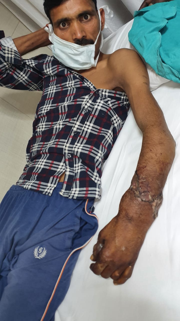
Changes in the blood, not the heart, may underlie cardiac thrombosis in COVID-19 patients

“My laboratory has a long history of defining endothelial cell alterations that produce pathologies, including thrombosis, and we expected to confirm the widely held assumption that local endothelial cell alterations were responsible for thrombosis of the cardiac vessels in COVID-19 patients,” explained lead investigator Jordan S. Pober, MD, PhD, Department of Immunobiology, Yale University School of Medicine, New Haven, CT, USA. “Instead, we found that the cardiac thrombi contained neutrophils that expressed changes known to promote coagulation, including changes that are associated with cell death and inflammation.”

Hospitalized patients with SARS-CoV-2 infection have an increased risk of developing myocardial injury. However, numerous studies have rarely detected viral protein or RNA within the hearts of patients who died from COVID-19, despite evidence of abundant virus presence in the lungs of the same patients. Thrombosis of micro and macro coronary vessels has most consistently characterized the hearts of individuals who succumbed to COVID-19, but the underlying cause remains unknown.

Dr. Pober and his colleagues examined heart tissue from seven autopsies of COVID-19 patients performed early in the pandemic, before anticoagulation treatment was commonly administered, and compared these specimens to autopsy tissue from 12 COVID-19-negative controls, with and without heart disease, using multiparameter fluorescence microscopy to analyze the composition of the thrombosed vessels. All patients in the COVID-19 group had severe pneumonia. One patient experienced a sudden cardiac arrest outside of the hospital, two patients developed sepsis, and one patient had recurrent acute leukemia with thrombocytopenia. The COVID-19-negative controls included six patients with pre-existing cardiac disease.

Thrombosis was the most common pathological finding in the COVID-19 group with a greatly elevated frequency of microthrombi and total number of macrothrombi compared to the COVID-19-negative controls. Despite the widespread evidence of thrombosis, no evidence of myocyte death or acute inflammation typically associated with myocardial infarction was detected in the COVID-19 group.

The vessels of the heart were examined for signs of endothelial cell injury, which can promote thrombosis through release of microparticles containing procoagulative tissue factor, or by endothelial cell sloughing that can expose platelet activating collagen. The investigators failed to find such endothelial changes at sites of thrombosis. Instead, they saw that the cardiac thrombi in four of the six COVID-19 patients contained neutrophils that expressed procoagulant changes in the blood, such as citrullination of histones associated with formation of neutrophil extracellular traps (NETs). Some images suggest NETS that appear to be directly associated with platelets. Neutrophil-rich macrothrombi composed of 30% or more neutrophils were common in the COVID-19 group but not in control tissue specimens.

Dr. Pober commented, “Our data challenge the view that alterations in the heart vessel wall are the primary cause of COVID-19 cardiac thrombosis. Current treatments of severe COVID-19 include anticoagulation, but the best strategy is still not clear. In light of our findings, reducing neutrophil responses could be an important target for therapeutic intervention. This and many other advances in the understanding of disease continue to be provided by autopsies, and I am grateful to the pathologists who performed them for this study at both Brigham and Women’s Hospital and Yale.”

Peter Libby, MD, a cardiologist and vascular biologist at Boston’s Brigham and Women’s Hospital and the Harvard Medical School, a long-time collaborator of Dr. Pober’s, stated: “For several years we have studied neutrophils and their prothrombotic products known as NETs in the context of clots that form in the larger coronary arteries. The finding of neutrophil involvement in the smaller blood vessels that course through the heart muscle in COVID-19 extends our understanding of cardiac injury that we often see in patients with severe SARS-CoV-2 infection. Brigham pathologists Robert F. Padera, Jr., MD, PhD, and Richard N. Mitchell, MD, PhD, helped us enormously by providing tissue samples for these analyses early on in our experience with this pandemic.”

Source: Elsevier

Medically Speaking

Share
Published by
Medically Speaking

Recent Posts

The Hidden Costs of Excessive Screen Time: How It Impacts Your Child’s Behavior and What You Can Do About It

Explore the profound effects of excessive screen time on your child’s behavior and development. Discover…

1 hour ago

Transform Your Heart Health: Simple Everyday Diet and Lifestyle Changes for a Happier, Stronger Heart

Discover how simple daily changes in your diet and lifestyle can significantly improve your heart…

1 hour ago

“Unraveling the Truth: Do Brain Tumours Only Affect Older Adults? Debunking Myths and Shedding Light on a Complex Condition”

This article explores the common misconception that brain tumours primarily affect older adults, revealing the…

2 hours ago

“Revolutionary Breakthrough: First-Ever Ovarian Cancer Vaccine Set to Transform Women’s Health and Combat a Silent Killer”

The first-ever ovarian cancer vaccine is poised to change the landscape of women's health by…

2 hours ago

“Battling the Monsoon Surge: Essential Strategies to Prevent Dengue and Chikungunya Infections in Delhi”

As dengue and chikungunya cases rise in Delhi during the monsoon season, understanding prevention strategies…

2 hours ago

NATURAL vs CHEMICAL SKINCARE: THE ESSENTIAL GUIDE TO CHOOSING WHAT’S BEST FOR YOUR SKIN

Skincare is a significant concern for women (and increasingly, men), with the quest for perfect,…

2 hours ago